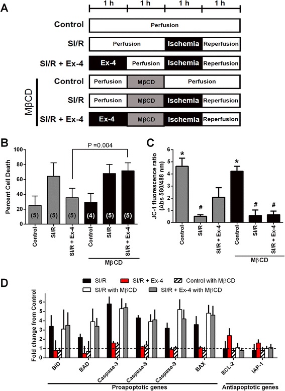
Figure 2.

In vitro assessment of the role of caveolae in exendin-4 (Ex-4) induced cardiac protection. (A) Summary illustration of in vitro experimental groups. (B) Cardiac myocytes exposed to simulated ischemia/reperfusion (SI/R) were exposed to experimental procedures outlined in A. Cell death was determined by trypan blue staining. Cardiac myocytes under control conditions (Control) had minimal cell death. Methyl-β-cyclodextrin (MβCD) abolished the Ex-4 induced cardiac protection effect. Group sizes are indicated on the individual bars in parentheses. (C) Apoptotic changes were measured by investigating mitochondrial membrane potential using JC-1 after SI/R. The excitation rate (red/green) indicates changes within the mitochondrial membrane potential. *P < 0.001 vs. SI/R, SIR + Ex-4, SI/R with MβCD, and SI/R + Ex-4 with MβCD. #P < 0.05 vs. Control, SI/R + Ex-4, and Control with MβCD. n = 4 per each group. (D) Real-time polymerase chain reaction analysis of pro-apoptotic and anti-apoptotic gene expression after re-oxygenation. n = 4 per each group.
