Figure 1.

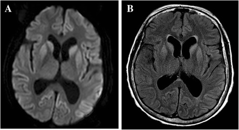
Patient MRI scans. Bilateral high signal intensities in the cortex, caudate nucleus, and putamen on DWI (A) and FLAIR (B).

Patient MRI scans. Bilateral high signal intensities in the cortex, caudate nucleus, and putamen on DWI (A) and FLAIR (B).