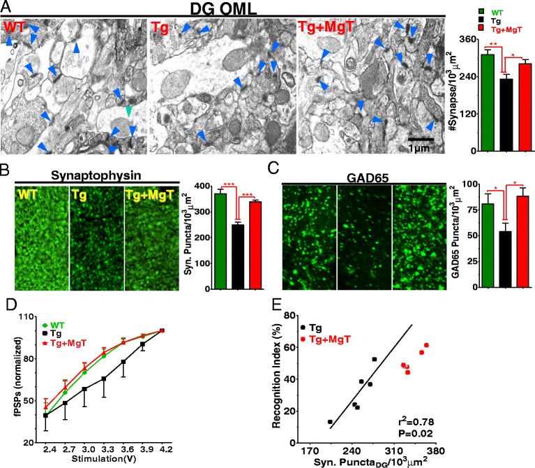
Figure 2.

Prevention by MgT treatment of synapse loss in APPswe/PS1dE9 transgenic mice (Tg mice). (A) Left: electron microscopic images showing structural synapses (blue arrows) in hippocampal outer molecular layer of dentate gyrus (DG-OML). Right: Estimated synaptic density. WT (n = 6), Tg (n = 6) and Tg + MgT (n = 5). (B) Left: Immunostaining of synaptophysin-positive terminals (Syn Puncta) in DG-OML. Right: Quantitative analysis of Syn Puncta (n = 6/group). (C) Same as in (B) and from same groups of mice; however, puncta represent GABAergic (GAD65). (D) Input–output (normalized) relationship of hippocampal CA1 synapses in vivo (n = 6/group). Field post synaptic potentials (fPSPs) were normalized by the maximum amplitude of fPSPs. Two Way ANOVA revealed significant effects of treatment: p < 0.05; and stimulus: p < 0.0001. (E) Correlation between the density of Syn Puncta and short-term recognition memory in Tg mice (23 months old). Tg + MgT (23 months old treated for 17 months) data are displayed but were not included in the regression analysis (Pearson’s test). Error bars show SEM. * p < 0.05, ** p < 0.01, *** p < 0.001.
