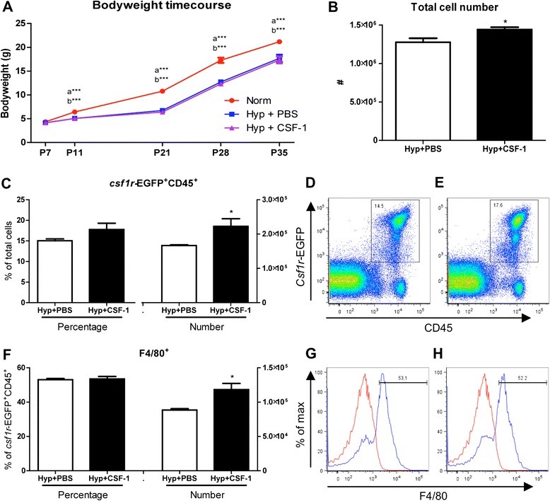
Figure 2.

Growth and macrophage analysis of mice treated with CSF-1 following hyperoxia. Body weights of mice treated with CSF-1 (Hyp + CSF-1; purple line) or PBS (Hyp + PBS; blue line) following hyperoxia exposure was significantly decreased compared to normoxic mice (Norm; red line) (A). n = 4-13 mice/time point. Data were analysed using one-way ANOVAs and Tukey’s post-hoc tests. ‘a’ represents significant difference between Norm and Hyp + PBS, ‘b’ represents significant difference between Norm and Hyp + CSF-1. Flow cytometric analysis of littermate lungs at P11 treated with CSF-1 or PBS controls following hyperoxia (B-H). Total lung cells (B) were gated on csf1r-EGFP+ CD45+ cells to quantitate CSF-1R+ cell number and proportion (C) after CSF-1 (D) and PBS treatment (E). F4/80 expression demarcates mature macrophages within the CSF-1R+ population (F-H). Histograms revealing gating procedure for F4/80 expression in representative PBS (G) and CSF-1-treated lungs (H). Staining (blue) is overlayed with an isotype control (red). CSF-1 treatment resulted in a significant increase in total cellularity, CSF-1R+ cell number and macrophage number. n = 4 littermate lungs/treatment. Data were analysed using two-tailed unpaired t-tests. *p < 0.05, ***p < 0.001.
