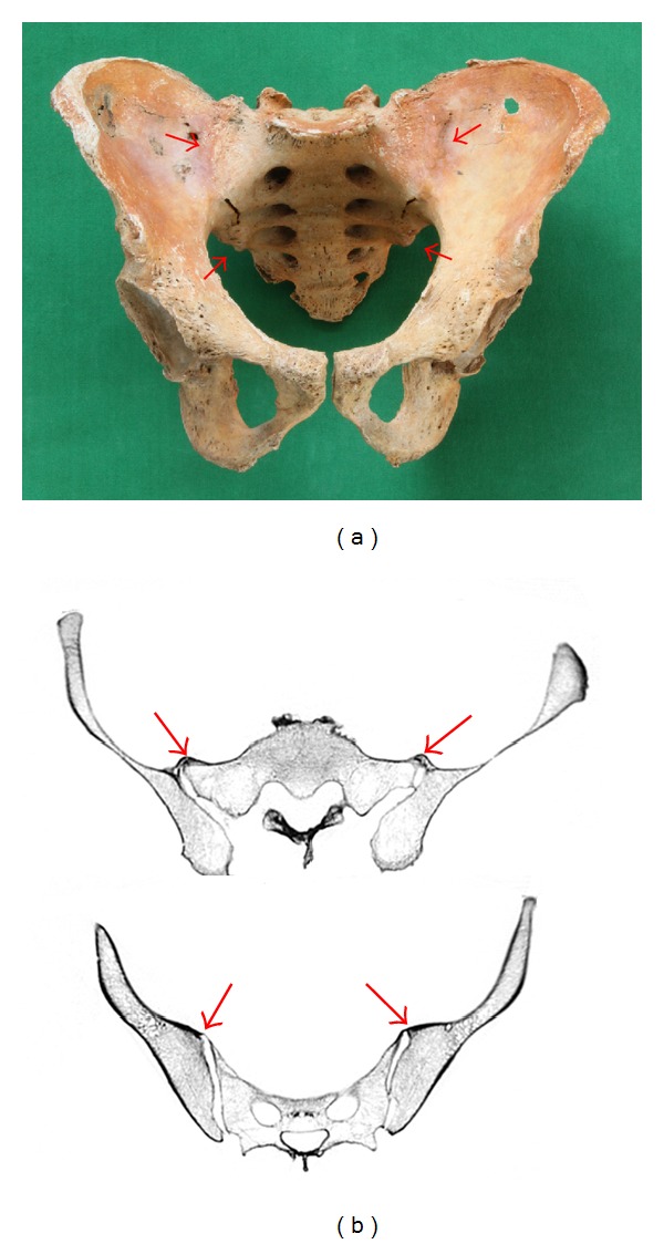
Figure 2.

(a) Pelvic bone of a 79-year-old male; both sacroiliac joints are fused with bony bridging. The sacroiliac joints of both sides are unioned in upper and lower portions (indicated by red arrows). (b) CT images of the same pelvic bone: bony bridging is localized in the surface area (indicated by red arrows).
