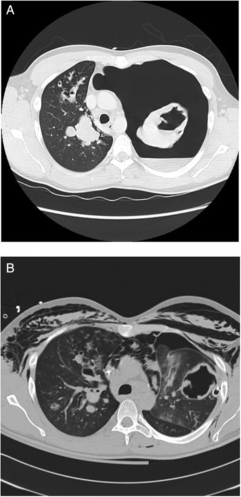
Figure 1.

(A) Initial chest CT demonstrating a complete left tension pneumothorax with an air-fluid level and a mediastinal shift, voluminous cavitations in the left lung, parenchymal infiltration in the right lung and mediastinal lymphadenopathy. (B) Chest CT 7 days after admission showing bilateral cystic bronchiectasis with mucoid impactions, multiple bilateral nodular opacities, mediastinal lymphadenopathy and persistence of voluminous cavitations in the left lung.
