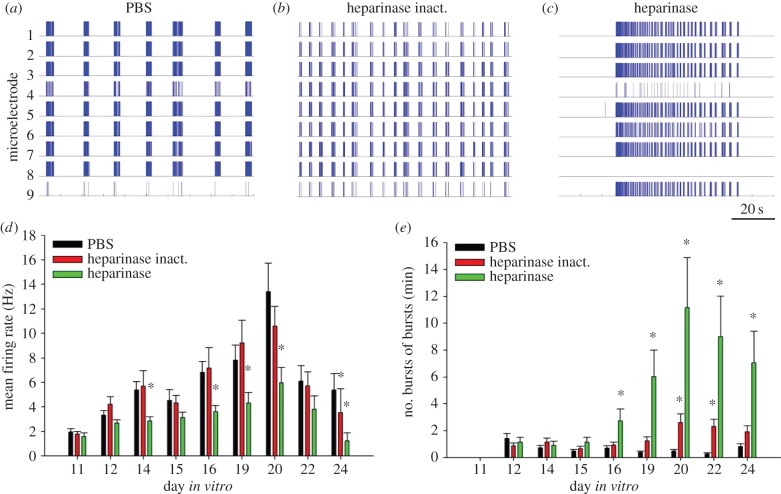
Figure 1.
Heparinase treatment induces epileptiform spiking activity. Raster plots of spontaneous spiking activity in primary hippocampal cultures at DIV 20, treated with PBS (a), heat-inactivated heparinase (b) or active heparinase (c). Scale bar is 20 s, valid for (a–c). Each stick of represented raster plots shows a spike recorded from one of nine electrodes of the microelectrode array. A high synchronization of activity recorded in most electrodes is obvious. The duration of demonstrated raster plots is 100 s. Neurons treated by heparinase show abnormally long (up to 70 s) epileptiform bursts of bursts, whereas cultures from control groups show shorter regular bursting episodes. A difference in bursting pattern in (a) and (b) reflects physiological variability, common for both control conditions. Developmental changes of mean firing rate (d) and mean frequency of bursts of bursts (e) in treated cultures. Both parameters of neuronal network activity reach the maximum at DIV 20. Increase of epileptiform activity in heparinase-treated cultures parallels the fall in overall mean firing rate relative to control groups. Data are presented as the mean ± s.e.m., n = 15–17. Statistical analysis was performed using a two-way analysis of variance (ANOVA), SNK was used as a post hoc ANOVA test. The difference between groups was considered significant if *p < 0.05. (Online version in colour.)

