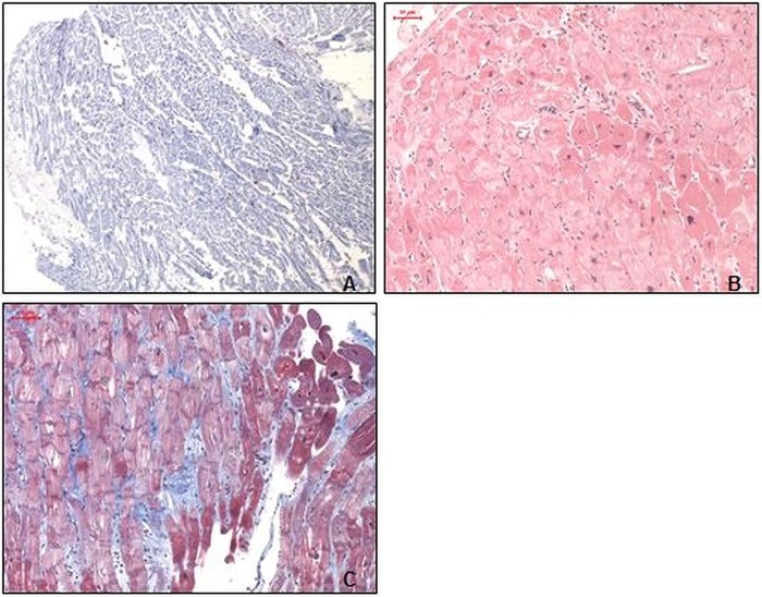
Figure 2.
Left ventricular endomyocardial biopsies after antithymocyte globulin treatment. (A) Normally distributed immunohistological staining of extensive macrophages (CD11b) infiltration (magnification ×100). (B) H&E staining with a largely inconspicuous myocardium, without infiltration of the endocardium (magnification ×100). (C) Azan staining with only a slightly increased extracellular matrix (blue; magnification ×100).

