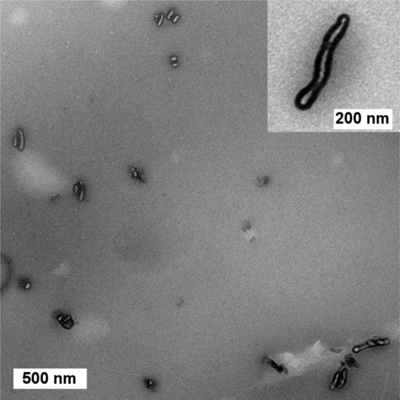
Figure 4.
TEM image of PS–PLA nanoparticles casted from a cyclohexane solution (0.02 g L–1) for 200 nm-thick sections. The particles were stained with RuO4. The TEM sample was prepared by absorbing the excess of solution with a piece of cleaning paper placed under the grid. Inset: A higher-magnification view of an isolated cylindrical nanoparticle revealing its core–shell structure.

