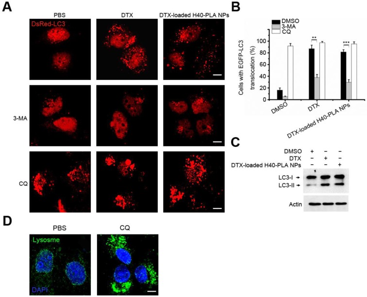
Figure 6.
CQ inhibited autophagy induced by DTX-H40-PLA NPs. (A, B) Representative images and quantification of MCF-7 cells with DsRed-LC3 vesicles. The DsRed-LC3 transfected MCF-7 cells were co-treated with 1 μM DTX and DTX-H40-PLA NPs for 24 h, respectively. Scale bars: 10 μm. Data are shown as the mean ± S.E.M. *P<0.05, ** P<0.01, *** P<0.001 compared to controls. (C) LC3I/II protein levels were analyzed by western blotting in the MCF-7 cells treated in (A). (D) MCF-7 cells were treated with 30 μM CQ for 24 h, the lysosome detected with LAMP1 antibody. Scale bars: 10 μm.

