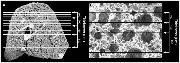
Figure 1.
Scanning electron microscopy (SEM) of the polymer casted murine lung. (A) Cross-sectional 12x SEM of murine lung including representative size measurements at right. Vascular fixation and casting – the method with the least shrinkage of common lung preparation techniques – demonstrated bronchiolar and alveolar duct architecture when examined by SEM. (B) 200x SEM image of murine lung demonstrating size of alveolar ducts. The alveolar ducts were demonstrably contained within the 200 μm tissue slice.

