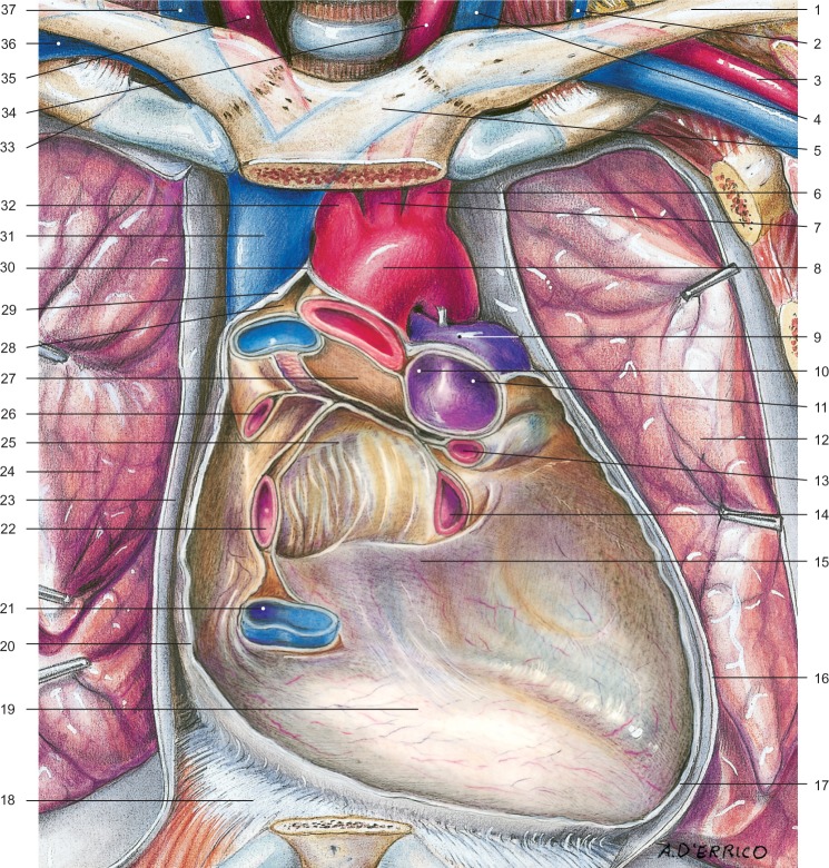
Figure 5.
Aspect of the pericardium after resection and removal of its anterior wall, the great vessels, and heart.
Notes: In evidence the base of the heart, the posterior wall and the areas of folding of the serous pericardium at the level of the great vessels. 1, left clavicle; 2, left external jugular vein; 3, left subclavian artery; 4, left internal jugular vein; 5, sternal manubrium; 6, left subclavian artery; 7, left common carotid artery; 8, carotid arch; 9, pulmonary trunk; 10, right pulmonary artery; 11, left pulmonary artery; 12, left lung; 13, left upper pulmonary vein; 14, left inferior pulmonary vein; 15, posterior wall; 16, left mediastinal pleura; 17, pericardial sac; 18, diaphragm; 19, base; 20, pericardial sac (dissected); 21, inferior vena cava; 22, right lower pulmonary vein; 23, right mediastinal pleura; 24, right lung; 25, diverticulum Haller; 26, upper right pulmonary vein; 27, transverse sinus of the pericardium; 28, visceral layer; 29, serous pericardium, parietal layer; 30, apex of the pericardial sac; 31, superior vena cava; 32, brachiocephalic trunk; 33, right first rib; 34, left common carotid artery; 35, right common artery; 36, right subclavian vein; 37, right internal jugular vein. Copyright Edi.Ermes, Milano. Reproduced with permission from Anastasi et al. AA VV, Anatomia dell’uomo, 4th ed, Edi.Ermes, Milano [Human anatomy].113

