Fig. 4.

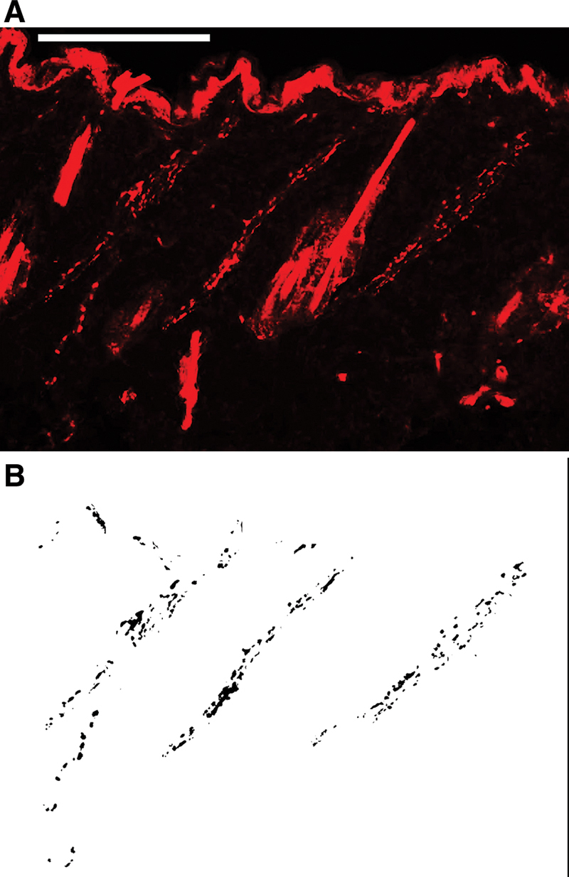
A, An image of PGP9.5 immunostaining of a section from unwounded skin. Scale bar: 100 µm. B, Images were analyzed using an automated method of quantifying the PGP9.5-positive area in each field of view with a digital image analysis program.

A, An image of PGP9.5 immunostaining of a section from unwounded skin. Scale bar: 100 µm. B, Images were analyzed using an automated method of quantifying the PGP9.5-positive area in each field of view with a digital image analysis program.