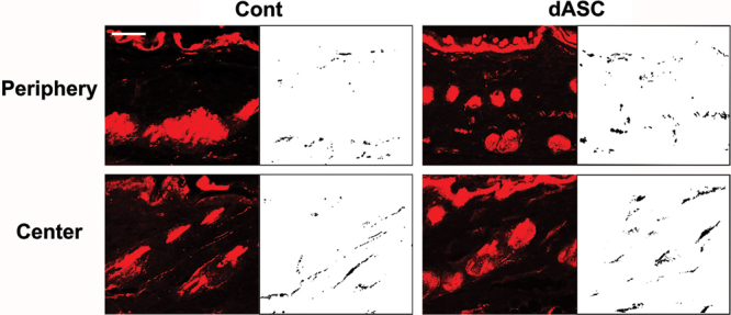
Fig. 6.

Images of PGP9.5 immunostainings of sections from innervated flaps. They were analyzed with a digital image analysis program. Note that axons tended to run parallel to the epidermis layer in the periphery of flap, whereas the innervation pattern in the center of flap was similar to that of unwounded skin. Scale bar: 50 µm.
