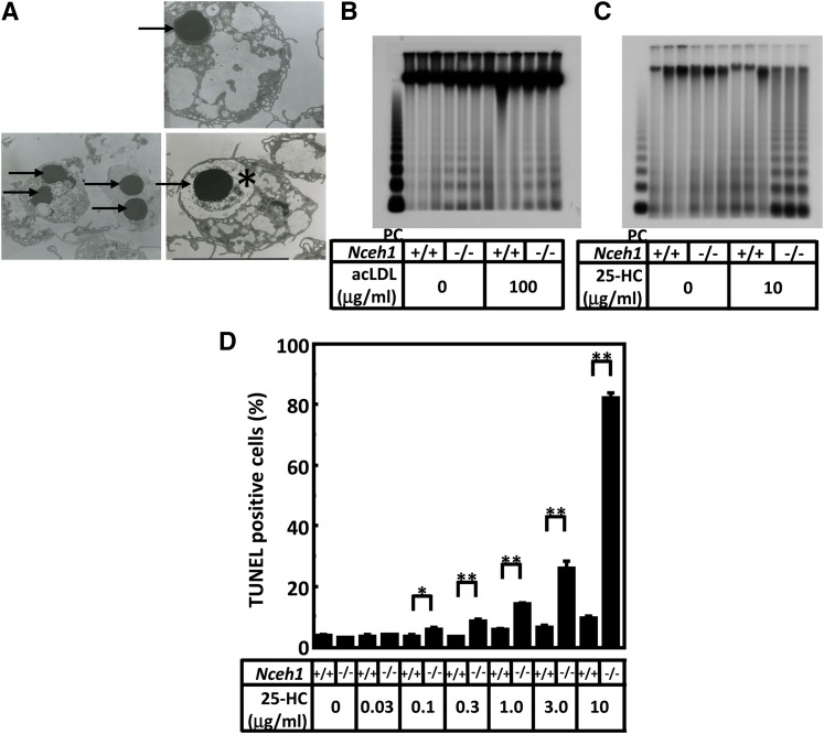
Fig. 1.
Nceh1-deficient TGEMs are prone to apoptosis. A: TEM observation. TGEMs were incubated in DMEM containing 10% FCS. Nuclear condensation (indicated by arrows), a characteristic feature of apoptotic cells, was observed predominantly in Nceh1-deficient TGEMs (representative images are shown). *The apoptotic macrophage engulfed by another macrophage. B, C: DNA ladder. DNA (0.5 μg) from macrophages was loaded in each lane. PC denotes positive control DNA (0.5 μg) from dexamethasone-treated thymocytes. Three wells of cells were incubated either in DMEM containing 10% FCS with or without acLDL (100 μg/ml) for 24 h (B) or in DMEM containing 10% LPDS with vehicle or 25-HC (10 μg/ml) for 24 h (C). D: Four wells of cells were incubated with increasingly higher concentrations of 25-HC for 24 h. The apoptotic cells were detected by TUNEL. Data are expressed as the mean ± SEM. *P < 0.05; **P < 0.01.

