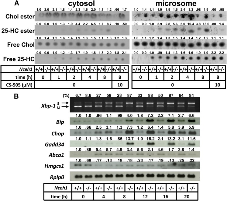
Fig. 3.
Involvement of activation of ER stress signaling is preceded by accumulation of 25-HC ester. A: TGEMs were incubated with 25-HC (10 μg/ml) for the indicated length of time in the presence or absence of CS-505 (10 μM). After cell fractionation, the lipids extracted from the cytosolic fraction (100 μg of protein) and microsomal fraction (50 μg of protein) were separated by TLC. Chol, cholesterol. B: Expression profile of ER stress markers. Cells were incubated with 25-HC (10 μg/ml) in the presence or absence of CS-505 (10 μM) for 12 h. s, spliced; u, unspliced. Values above each band/spot indicate the fold difference evaluated by densitometry. Xbp-1 splicing was assessed as the percent spliced Xbp-1[spliced Xbp-1/(spliced Xbp-1 plus unspliced Xbp-1)].

