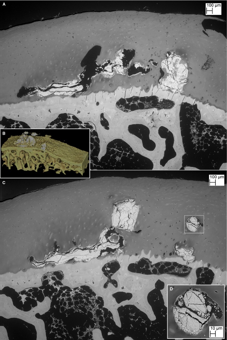
Fig. 2.

(A) BSE SEM of triiodide-stained PMMA block surface 260 μm further into the block than in Fig. 1G. This still shows the higher density of the HDMP, but the non-mineralised HAC can now also be visualised via the iodine staining. (B) Same level shown in drishti reconstruction from 6 μm μCT40 XMT dataset. (C) BSE SEM, finished 280 μm into the block, again after triiodide staining, shows HDMP surrounded by HAC. Also note region with no SCB under the ACC in the right half of the field. (D) Higher magnification showing detail from white box in right hand of panel (C). Note cracking to produce fine fragments and sharp edges.
