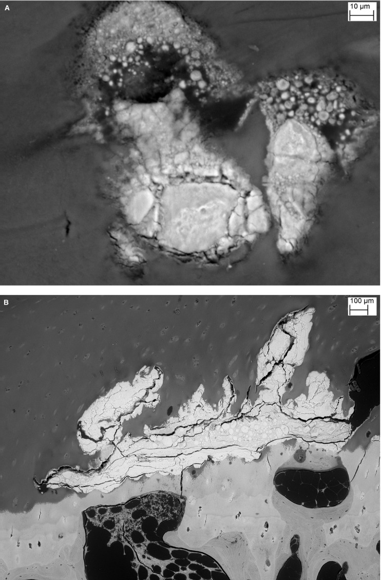
Fig. 4.

(A) Higher magnification of area outlined with white box in Fig. 3A showing detail of the mineralisation pattern within the HDMP with partial fusion of spherical mineralisation clusters. (B) Surface finished 550 μm into the block, triiodide staining, shows HDMP surrounded by HAC above with parts confluent with bone and ACC below. No SCB under ACC to left of centre. Chondrocytic lacunae in the ACC are mineralised.
