
Fig. 3.
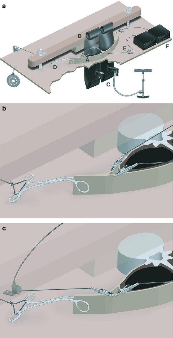
(a) Experimental apparatus design. Cadaveric slab (A) is placed on a wooden platform with holes to accommodate inflatable tubes (C) attached to a positive displacement pump (note: jagged cut-out is to demonstrate spatial context). To prevent vertebral rotation, a cross-bar (B) is placed across the vertebral body and clamped down using wingnuts on threaded bolts. A hemostatic clamp (D) attaches the common aponeurosis of the transversus abdominis and internal oblique muscles the CTrA to a constant load. Alligator clips attach the anterior and posterior laminae of the common aponeurosis to load cells (E) that connect to load cell meters with digital display (F). (b) Experimental apparatus design. Magnified view of alligator clip placement for differentiating MLF/PLF force transfer (Method 5). Alligator clips are attaching the anterior and posterior laminae of the common aponeurosis to load cells (not shown). Hemostatic clamp is attaching the common aponeurosis to a known load (not shown). For simplicity, the inflatable tubes are not shown in this image. (c) Experimental apparatus design. Magnified view of alligator clip placement for analyzing the effect of inflation on PLF force transfer (Method 6). Alligator clips are attached along the posterior laminae of the common aponeurosis in opposing directions. Hemostatic clamp is attaching the common aponeurosis to a known load (not shown). For simplicity, the inflatable tubes are not shown in this image.