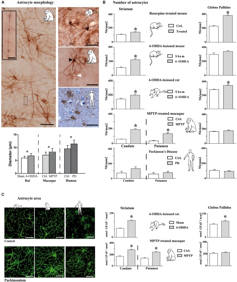
Figure 2.
Morphological and quantitative analysis of astrocytic reaction to dopamine depletion in striatum and GP of animal models of PD and parkinsonian patients. (A) GFAP-S100β immunostaining enables demonstrating significant increase in astrocyte soma diameter in the rat and monkey models of PD (n = 4 sham and 4 6-OHDA rats; n = 3 control and 3 MPTP monkeys; mean ± SEM) (scale bar = 30 μm but in inset, 10 μm). Primates show two level of immunoreactivity indicated by a black arrowhead for intense labeling and a white arrowhead for moderate staining. (B) Quantitative analysis of astrocyte number in striatum and GP in controls and in animal models of PD (n = 4 reserpine mouse, n = 5 6-OHDA mouse, n = 4 6-OHDA rat, n = 3 MPTP non-human primate) as well as in parkinsonian patients (n = 3) showing increase in astrocyte number. * denotes a significant difference (Student t-test; p < 0.05). (C) Area analysis of astrocytic processes after GFAP immunostaining and 3D reconstruction. Left: representative examples of GFAP immunostaining in the striatum of 6-OHDA rat and MPTP monkey models of PD as well as in PD patients (Scale bar = 30 μm). Right: Astrocytic area significantly increases in the striatum of 6-OHDA rat (n = 4) and MPTP monkey (n = 3) models of PD. * denotes a significant difference (Student t-test ; p < 0.05).

