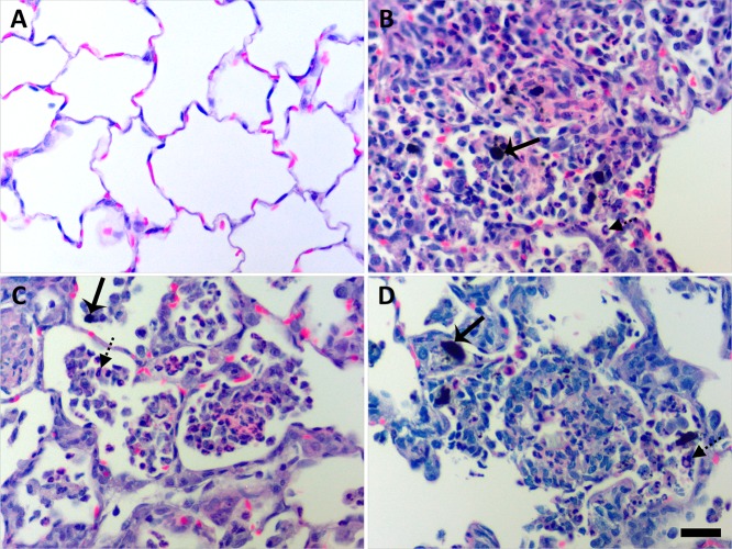
Figure 9.
Instilled MWCNTs produced particle-associated inflammation in the lung parenchyma at day 1. Histopathological findings from exposure to DM (A), P-MWCNTs (B), O-MWCNTs (C), and F-MWCNTs (D). Panels are bright-field microscopy images of the most severe responses observed in lung tissues from rats given a single 200 μg dose of MWCNTs or 250 μL of DM. Tissues were stained with H & E. Solid arrow = MWCNTs, and broken arrow = inflammatory polymorphonuclear cell. Scale bar (25 μm) applies to all panels.

