
Figure 2.
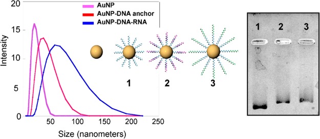
Size determination of ligated siRNA SNAs. (a) DLS data showing the increase in size between the citrate-capped 13 nm Au nanoparticles (pink), the SNAs modified with the DNA ligation anchor (red) and the final RNA-DNA chimera formed at the surface of the Au NPs (blue). (b) Electrophoretic mobility of the ligated siRNA SNA and its intermediates. DNA anchor functionalized SNA (lane one), DNA anchor modified SNA hybridized to the RNA specific DNA bridge (lane 2), fully ligated siRNA construct post treatment with T4 DNA ligase and removal of DNA bridge (lane 3).