
Figure 3.
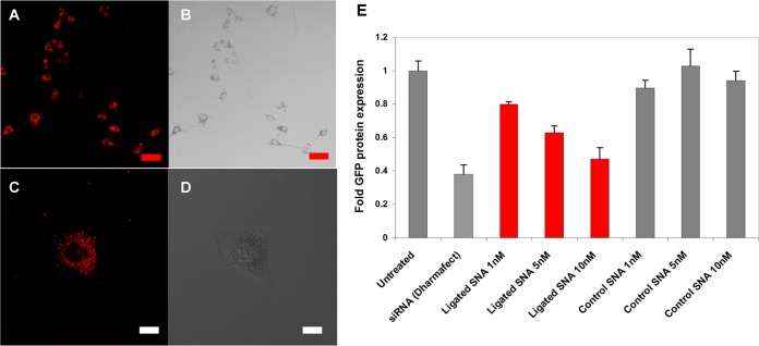
Cellular uptake and gene knockdown of GFP by siRNA ligated SNAs. (a) Confocal microscopy of C166 cells treated with 5 nM Cy5-labeled SNAs. (b) Same image as (a) without Cy5 filter. (c) Individual cell showing the diffuse uptake of the ligated SNAs into the cell as indicated by Cy5 fluorescence. (d) Corresponding bright field image of cell shown in (c). (e) Comparison of C166 GFP-expressing cells by flow cytometry showing relative changes in the amount of GFP protein expression post treatment with GFP siRNA ligated SNAs. Compared to the untreated cells, 10 nM treatment of the cells with the ligated SNAs resulted in 53% decrease in GFP expression. The ligated SNAs also follow a dose-dependent knockdown of GFP protein over the concentrations investigated. Scale bar is 50 μm in a, b and 10 μm in c, d.