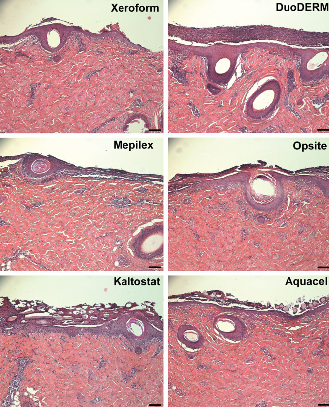
Fig. 4.

Representative hematoxylin-eosin-stained sections of wounds 3 d after treatment. Mepilex-treated wounds showed less epithelial migration extending outward from hair follicles than the other dressings. A mixture of fibrin clot, dead polymorphonuclear leukocytes, and degraded extracellular matrix components at the interface between the dressings and the healthy underlying dermis can be visualized sloughing off after epidermis formation. Note Kaltostat and Aquacel fibers surrounded by the mixture of the dermis-clot interface. Scale bar = 100 μm.
