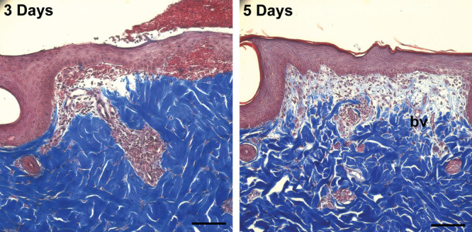
Fig. 7.

Representative Masson trichrome–stained sections of wounds treated with DuoDERM on days 3 and 5 after surgery demonstrating granuloma tissue formation and tissue remodeling. By 5 d, the number of inflammatory cells has decreased and the number of fibroblasts has increased, with new collagen (light blue) and blood vessel (bv) formation visible. In addition, the epithelium is maturing with stratum corneum observed. Scale bar = 100 μm.
