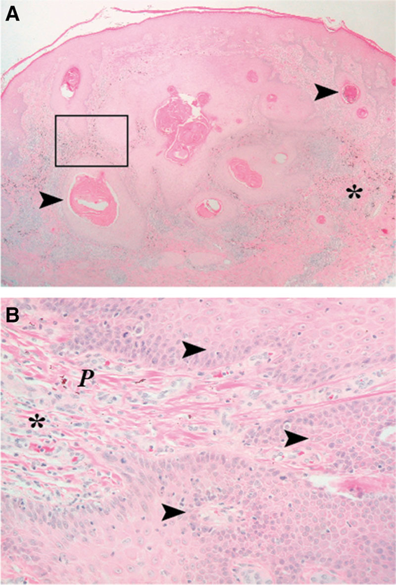
Fig. 1.

Histopathological verification of a SCC. A, As a relative well-circumscribed neoplasm in the dermis, the tumor is composed of strands and nodules of atypical squamous cells with some mitoses and furthermore shows endophytic prolongation into the dermis. Also variable central keratinization and horn pearls formation can be found. Magnified rectangular cutout shown in (B) with typical nests of squamous cells alongside with atypia and mitotic activity in the dermis, near to the tumor formation red pigment, can be seen. Arrow, SCC complex; P, red exogenous pigment; *, infiltrate of histiocytes and lymphocytes; (A), 25-fold microscopic magnification, and (B), 200-fold microscopic magnification.
