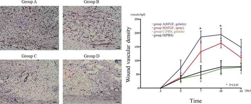
Fig. 4.

Wound vascularization. A, Anti-CD31 antibody staining at day 7. The brown-colored areas correspond to blood vessels stained by the anti-CD31 antibody. Stronger CD31 staining was observed for group A compared with the other groups, indicative of a larger number of vascular structures. B, Graph demonstrating density (pixels/hpf) of CD31-positive staining over the time course of 3–14 d. Group A showed a significantly higher number of CD31-positive pixels compared with the other groups at day 7 and day 10 (bar: 50 µm).
