
Fig. 2.
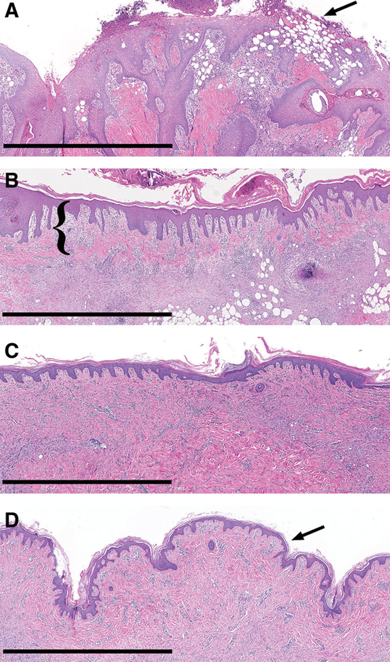
Histology (H&E) of wounds treated with MSTCs (A, C) or STSG (B, D), at 1 (A, B) and 5 wk (C, D) after treatment. One week after treatment, wounds treated with MSTCs were partially reepithelialized, with intermittent gaps still remaining in the new epithelial layer (A, arrow) at the wound surface. By contrast, STSG provided immediate coverage (B, STSG highlighted by bracket), with granulation tissue developing under the graft. By 2 wk after treatment, MSTC-treated wounds were also completely reepithelialized, and epidermal remnants were no longer seen in the dermal region (not shown). The uneven surface that is characteristic of meshed STSGs were also evident in histology as prominent folds in the epidermal and upper dermal layers (D, arrow), and these persisted through the course of the experiment. Scale bars: 2 mm.