
Fig. 1.
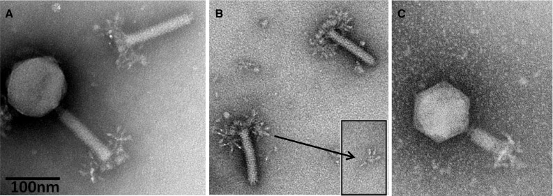
Electron micrographs of Salmonella phage ViI. (A) Complete phage ViI with quiescent tail and severed tail; (B) ViI tails with unfolded tail entities, displaying an umbrella-like structure; (C) ViI with contracted tail. The phage was applied to a glow-discharged carbon/Formavar-coated 200-mesh copper grid and then stained with 5 % aqueous ammonium molybdate plus 1 % trehalose. The grid was finally examined on a 120 KV Philips Tecnai Spirit BioTwin transmission electron microscope fitted with a Tietz F415 charge-coupled-device (CCD) TemCam camera