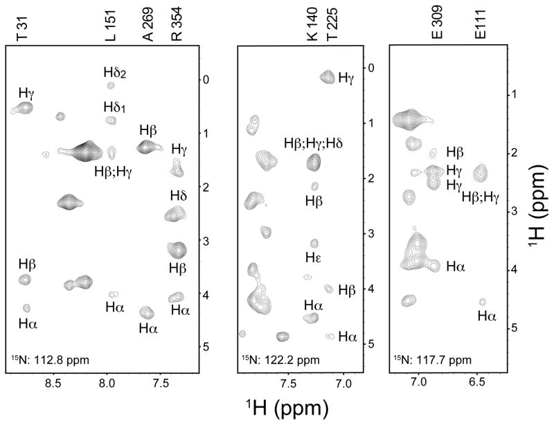
Figure 6.
15N planes of a H(CC)(CO)NH TOCSY spectrum of uniformly 15N,13C-labeled MBP in CTAB/hexanol reverse micelles dissolved in liquid ethane at 4,500 psi. The 42 kDa monomeric protein was 0.2 mM in 75 mM CTAB and 450 mM d-hexanol reverse micelles with a water loading of 15. The sample was prepared from a stock solution of 5 mM MBP in 20 mM sodium phosphate, pH 7.5, with 5 mM EDTA and 7 mM β-cyclodextrin. The spectrum was acquired at 600 MHz (1H) at 20 °C using a cryoprobe. The data set was obtained using 128 scans per FID and included 24 complex increments in the nitrogen dimension, 42 complex increments in indirect hydrogen dimension. The DIPSI carbon TOCSY mixing period was 16 ms. Various 15N planes are shown to illustrate the richness of the remote correlations obtained. Indicated assignments were mapped from those obtained using a triple resonance assisted main chain directed assignment strategy (Xu et al. 2006).

