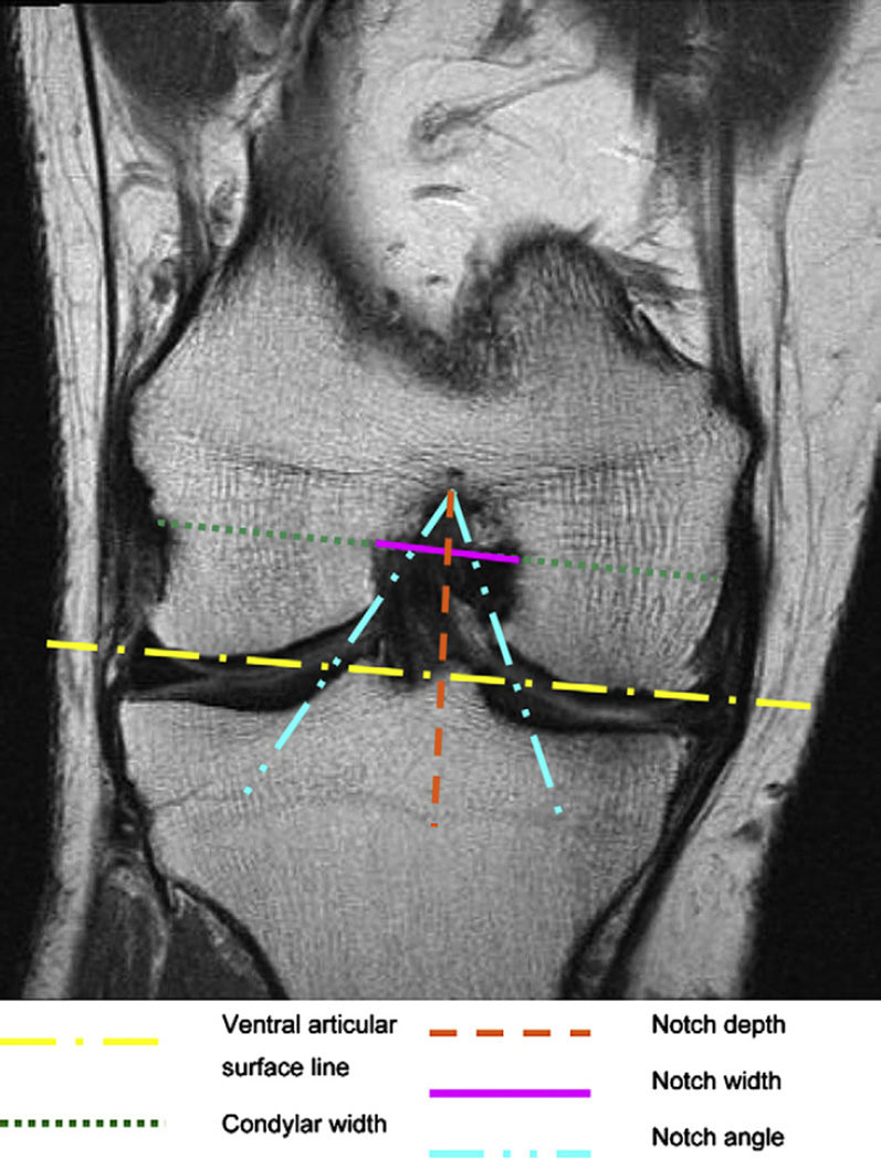
Fig. 3.
Measurements of the femoral notch on a coronal image. The notch depth is perpendicular to a line connecting the ventral articular surfaces of the medial and the lateral femur condyle. The notch width and the condylar width are parallel to that line at 2/3 of the notch depth. MR details: coronal IW TSE (COR_IW_TSE), slice thickness of 3 mm, 3850 ms TR, 29 ms TE, 180° FA, 14 cm × 14 cm FOV; 384 × 307 pixels matrix; 352 Hz/pixel bandwidth; acquisition time 3 min 24 s.

