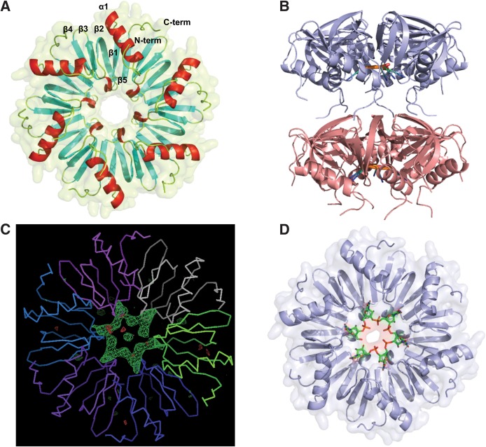
FIGURE 1.
Structures of Lm Hfq and the Lm Hfq-U6 complex. (A) View into the proximal face of the Lm Hfq hexamer. The protein is shown as a cartoon and surface. The N and C termini of the protein are labeled, while the α helices are colored red, and the five-stranded twisted antiparallel β sheet is shown in cyan. The secondary elements of one protomer are labeled. (B) The asymmetric unit of the Lm Hfq–U6 complex structure. Notice the N terminus-distal face packing. The Lm Hfq is shown as a blue cartoon and the bound RNA as an atom-colored cartoon. (C) An Fo – Fc electron density difference map calculated after molecular replacement and one round of rigid body refinement in Phenix and prior to the addition of the U6 RNA. The Cα trace of the protein backbone is shown with each protomer individually colored. The electron density difference map (green mesh) is contoured at 3.5 σ. (D) View into the proximal face of the Lm Hfq–U6 complex structure. The protein is shown as a cartoon and surface colored blue, with the RNA shown as sticks and colored by atom type, with carbon atoms colored green.

