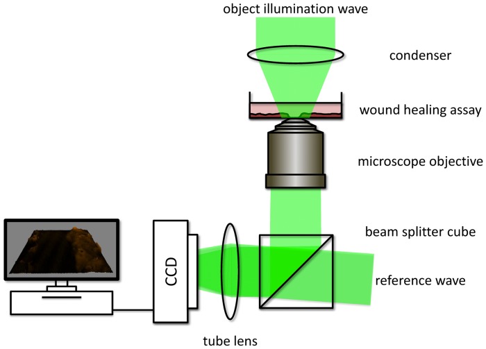
Figure 1. Utilized off-axis setup for digital holographic microscopy (DHM).
A laser beam is divided by a beam splitter into an object wave, illuminating the specimen through a condenser and an undisturbed reference wave. The object wave interferes with the slightly tilted reference wave on a charge coupled device sensor (off-axis geometry). Morphological changes of the biological specimen lead to changes of the optical path length of the object wave, which are coded in the resulting interference pattern (digital hologram).

