
Fig. 1.
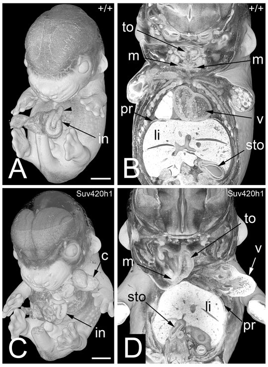
Multiple defects in a TS21+ mouse embryo (Suv420h1−/−). 3D volume rendered models. (A) External aspect of a wild-type (+/+) embryo. Note the intestine (in) forming the umbilical hernia and the caecum (arrowhead). (B) Coronally sectioned 3D model of this embryo. Note the mandibles (m) in the left and right lower jaw, the symmetric appearance of the tongue (to), the diaphragm (pr) cranial to the liver, and the position of the organs. (C) External aspect of a Suv420h1−/− embryo. Note the thoracoschisis, the position of the heart (c), and the arrangement of the intestine. (D) Coronally sectioned 3D model of this embryo. Note the following malformations: the left lower jaw is missing. The tongue is thin and fixed only on the right mandible. Cardiac ventricles (v) are outside the body cavity. Stomach (sto) is on the right side. The liver (li) is shifted cranially into the thoracal cavity. Scale bars: 1 mm.