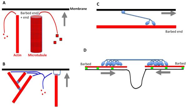
Figure 2. Mechanisms for cytoskeleton-based force generation on membranes.
Membrane in black, cytoskeletal element in red, cytoskeletal-interacting protein in blue. Gray arrows denote direction of membrane movement.
LEFT: Polymerization-based force generation. A) Membrane can be pushed by a polymerizing actin filament or microtubule by new polymer subunits adding to the + end (barbed end for actin), which abuts the membrane. B) Specific examples of actin polymerization-based force production. Arp2/3 complex (left) assembling a branched filament network (Arp2/3 complex is at the branches), or a formin protein (right) attached to the filament barbed end and to the membrane. Not shown are proteins containing multiple WH2 domains (Spire, Cordon Bleu), that can nucleate new actin filaments, possibly remaining at barbed ends in some cases.
RIGHT: motor-based force generation. C) Motor-based translocation along a filament. Dimeric myosin motor moving towards the actin filament barbed end using its motor head groups, and attached to a membrane through a tail motif. One myosin (myosin VI) moves toward the opposite end (the pointed end). Motors that translocate on microtubules include kinesins (toward plus end) and dyneins (toward minus ends). D) Motor-based contraction. A non-muscle myosin II mini-filament, in which the motor heads are bound to two different actin filaments, and moving towards their respective barbed ends. In the process, the myosin II causes constriction of the membrane attached to the actin filaments.

