Fig. 3.
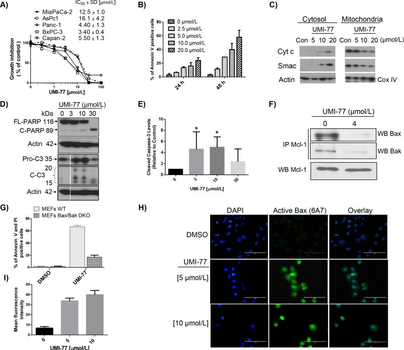
Fig. 3. 2 (UMI-77) effect on pancreatic cancer cell growth and induction of apoptosis
A) IC50 values of cell growth inhibition of 2 (UMI-77) in panel of PC cell lines after 4 days treatment. B) Time and dose-dependent induction of apoptosis in Panc-1 cells after treatment with 2 (UMI-77). Cells were treated for different time points and apoptosis was determined with Annexin V/PI double staining. C) Release of cytochrome c and Smac from mitochondria in Panc-1 cells. Cells were treated for 24 h, mitochondria were isolated and cytochrome c and Smac were probed by Western blotting. D) Full length PARP (FL-PARP), cleaved PARP and activation of caspase-3 in BxPC-3 cells after 24 h treatment with 2 (UMI-77). Cells were treated for 24 h, and caspase-3 and PARP were probed by Western blotting. E) Cleaved caspase-3 was quantified by densitometric analysis and presented in graphical form. The statistical significance was calculated with minimum of three values for all tested concentrations (n=3; P < 0.05). F) Immunoprecipitation on 2 (UMI-77) treated BxPC-3 cell lysate was performed using Mcl-1 antibody followed by western blot analysis with Bax or Bak. G) Induction of apoptosis in wild type (WT) and Bax/Bak-deficient (DKO) MEFs cells after 24 h treatment with 10 μM of 2 (UMI-77). H) and I) 2 (UMI-77) induces Bax activation in Panc-1 cells. Immunocytochemistry analysis (H) and quantification of the fluorescence intensity (I) demonstrated the increased number of positive cells stained with anti-Bax(6A7) antibody, which specifically detect the active form of Bax, 24 h after 2 (UMI-77) treatment in Panc-1 cells. Conversely, DMSO control did not induce the active form of Bax (green: anti-Bax(6A7) antibody, blue: DAPI). Scale bar, 100 μm. Forty cells were used for quantification of the fluorescence intensity. *All experiments were performed minimum three times and the most representative results are presented.

