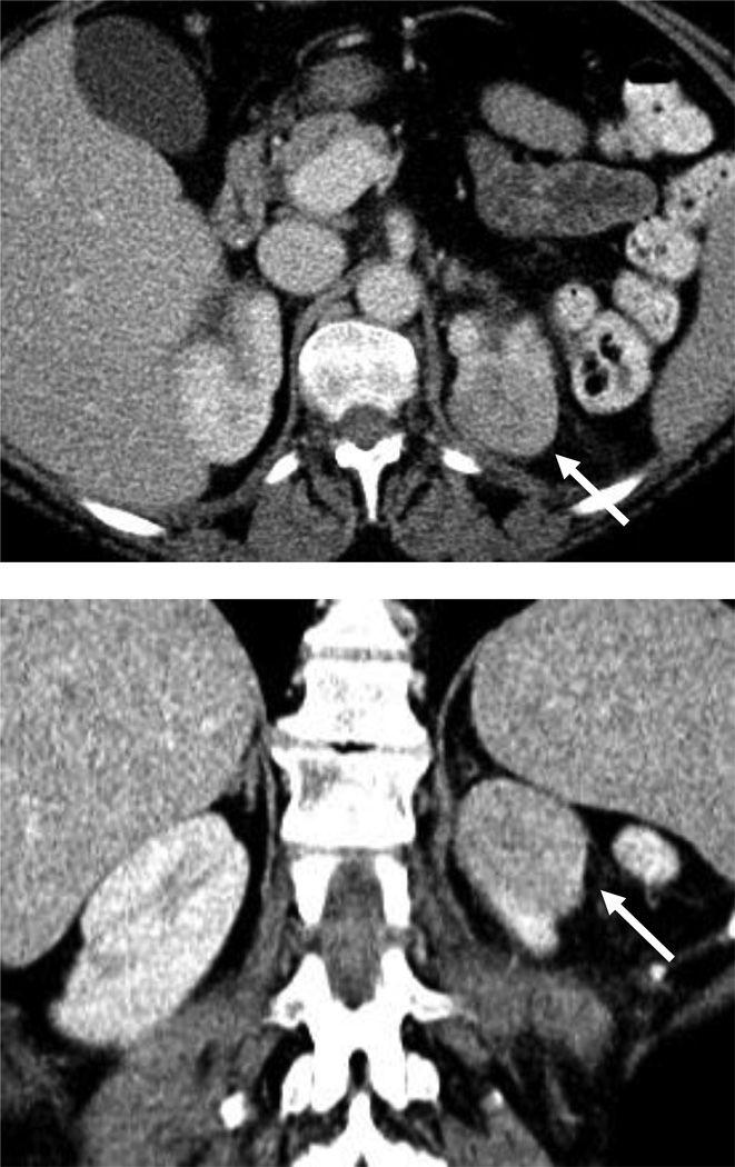
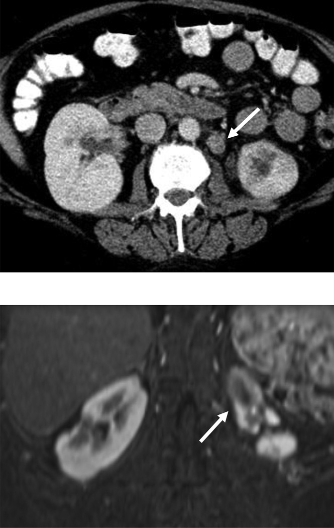
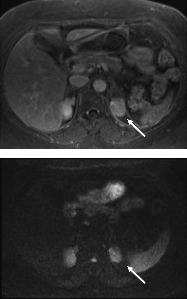
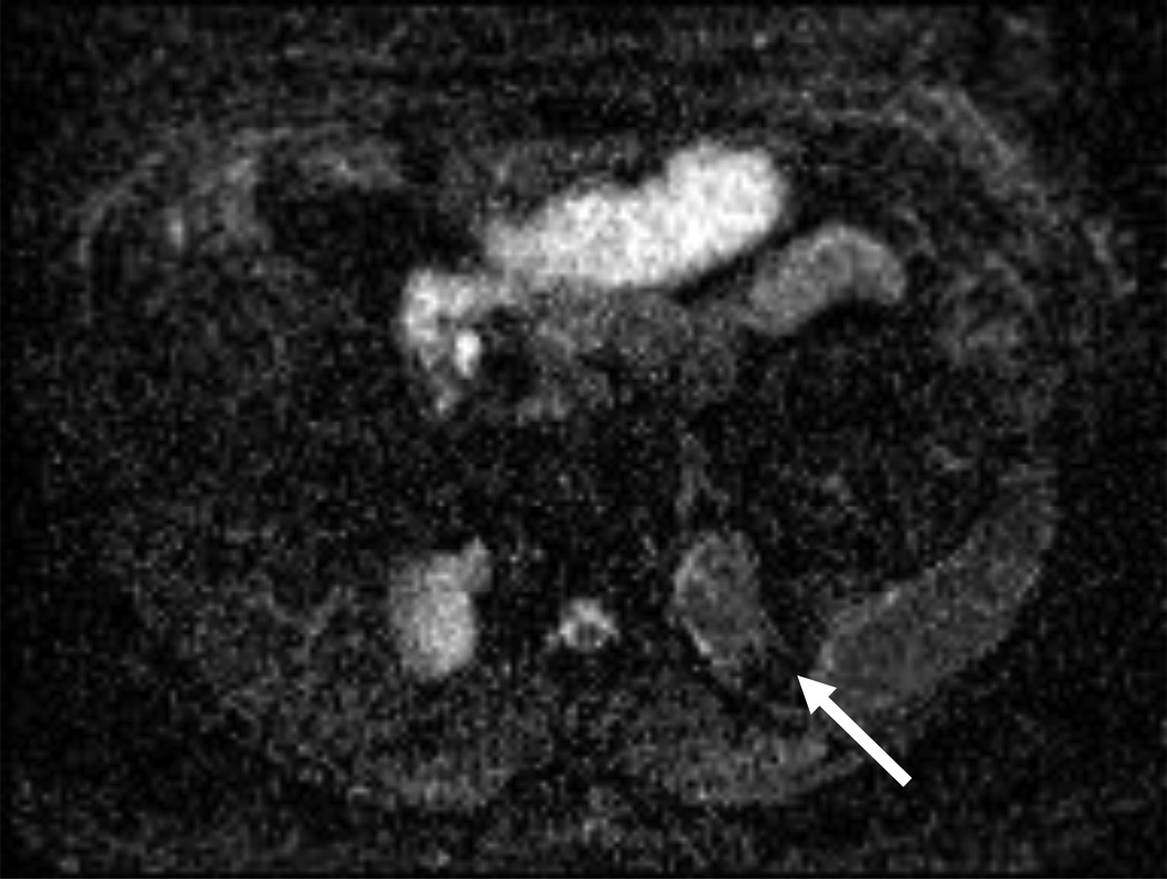
Figure 3.
54 year-old woman with a left renal mass at CT and clinical diagnosis of pyelonephritis.
A and B, CT with and without contrast shows apparent left upper pole mass with hypoenhancement relative to renal parenchyma. C, Enlarged retroperitoneal lymph nodes are also present.
D and E, Dynamic contrast-enhanced MRI one week later shows slightly smaller lesion, with corticomedullary differentiation internally, and continued hypoenhancement relative to parenchyma in the nephrographic phase.
F and G, Lesion also demonstrates marked restricted diffusion with loss of signal on corresponding ADC map. Focal pyelonephritis was diagnosed and resolved at follow-up MRI.


