
Figure 7.
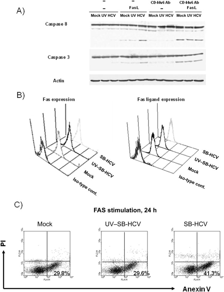
Susceptibility of different cells to Fas stimulation. A, Immunoblotting analysis was carried out by using anti-caspase 8, anti-caspase 3, and anti-actin antibodies, and the membrane was then subjected to reaction with peroxidase-conjugated secondary antibody. Immunoreactivity was visualized by use of enhanced chemiluminescence detection. Loading proteins were obtained from the following cell groups: mock-infected Molt-4 cells; Molt-4 cells infected with UV-irradiated hepatitis C virus, strain B (UV–SB-HCV); SB-HCV–infected Molt-4 cells from the carboxyfluorescein succinimidyl ester (CFSE)–high group; and SB-HCV–infected Molt-4 cells from the CFSE-low group, with or without Fas-ligand (FasL) stimulation. Pretreatment with CD44 splicing variant 6–blocking antibody (CD44v6 Ab) was carried out for some samples, as indicated. B, Surface expression of Fas and FasL as measured by flow cytometry. C, Fluorescence-activated cell sorter analysis of annexin V and propidium iodide (PI) staining. Mock-infected cells, Molt-4 cells infected with UV-SB-HCV, and Molt-4 cells infected with SB-HCV were stimulated with FasL for 24 h and analyzed to detect annexin V–positive, PI–negative early apoptotic cells. The percentages of early apoptotic cells in each group are indicated in the dot plots. Iso-type cont., isotype control antibodies.