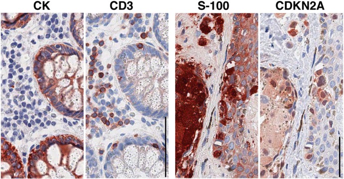
Figure 5.
Routinely processed sections sequentially immunostained after elution and re-staining with diagnostic antibodies. One section of normal colon (left two images) and one section from a cutaneous nevus (right) are stained sequentially for the antigens named above each image. The primary antibodies have been counterstained with a multilink (mouse + rabbit) HRP-conjugated polymer. Note the clean background and no crossover staining left by the earlier staining. Bar = 100 µm.

