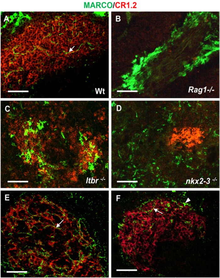
Figure 3.
Follicular display of MARCO requires mature follicular dendritic cells (FDCs) and marginal zone (MZ). Splenic sections from wild-type spleen (A) or from mice with various mutations (indicated in each micrograph, B–D) that affect the splenic architecture are shown. The association between CR1/2 (red) and MARCO (green) in WT spleen (A) is indicated (arrow). Mice with blocked lymphoid development (RAG1-deficient mice, B) lack mature FDCs expressing CR1/2 and follicular MARCO+ conduits, but contain MZ macrophages. Mice deficient for LTβR (C) have disorganized white pulp and MZ structure, and lack white pulp-associated MARCO. Nkx2-3-deficient mice (D) are unable to establish follicular MARCO-positive conduits in the absence of MARCO-positive MZ macrophages (n=5 from each genotype). (E) Clodronate liposome-mediated depletion of MZ macrophages efficiently depleted the producer cells within 48 hr, whereas the follicle-associated MARCO persisted in a close association with FDCs. (F) Ten days later, both follicle-associated MARCO and partial MZ repopulation by MARCO-positive MZ macrophages can be observed (n=6). Arrows in A, E and F point to follicular MARCO; arrowhead in F indicates a MARCO-expressing MZ macrophage. Bar size, 50 µm.

