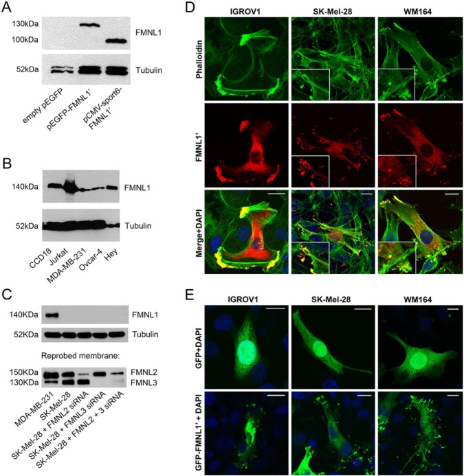
Figure 2.
Detection of the FMNL1’ construct and endogenous FMNL1 by western blotting and fluorescence microscopy. (A) HEK 293T cells were transfected with pCMVsport6-FMNL1’, pEGFP-FMNL1’ or empty pEGFP expression constructs. In control cells transfected with empty pEGFP, no FMNL1 band was detected in western blotting. In transfected cells, a 130-kDa band corresponding to GFP-tagged FMNL1’, and a 100-kDa band corresponding to non-tagged FMNL1’ is visible. (B) Endogenous FMNL1 is detected as a single band of 140 kDa in several cell lines: myofibroblasts (CCD18), T-cell lymphoma (Jurkat), breast cancer (MDA-MB-231) and ovarian cancer (Ovcar-4 and Hey). (C) To confirm that the FMNL1 antibody does not cross-react with FMNL2 or FMNL3, cell lysates were first blotted with the FMNL1 antibody, and reprobed with an antibody that cross-reacts with both FMNL2 and FMNL3. In MDA-MB-231 cells, a single band corresponding to FMNL1 was visible in MDA-MB-231 cells but not in SK-Mel-28 melanoma cells. In both cell lines, bands corresponding to FMNL2 and FMNL3 were present, indicating that the FMNL1 antibody specifically detects FMNL1, not FMNL2 or FMNL3. The specificity of the FMNL2 and FMNL3 antibody was further confirmed by reduced reactivity after corresponding siRNA treatments. Tubulin was blotted as a loading control. (D) IGROV1 ovarian cancer cells and the WM164 and SK-Mel-28 melanoma cell lines, which do not express endogenous FMNL1, were transfected with an FMNL1’ construct and stained with green phalloidin to visualize actin filaments (upper panel) and FMNL1 (red, middle panel). In membrane protrusions, FMNL1’ co-localizes with bundled F-actin (merge in yellow, bottom panel). Higher magnification of membrane protrusions are depicted in insets. (E) IGROV1, WM164, and SK-Mel-28 cells were transfected with a construct expressing GFP-FMNL1’ or GFP only. GFP shows a diffuse cytoplasmic distribution (upper panel), whereas GFP-tagged FMNL1’ is partly located at the membrane protrusions. Scale bars, 20 μm. Nuclei were stained with DAPI (blue).

