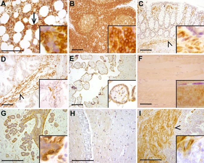
Figure 4.
Distribution of FMNL1 in human tissues. (A) In the bone marrow, megakaryocytes and erythroid cells do not express FMNL1, whereas myeloid cells stain strongly (arrow). (B) In lymph nodes, mature lymphocytes stain strongly. (C) In the colon, epithelial cells do not express FMNL1, whereas stromal lymphocytes and smooth muscle show strong reactivity (arrowhead). (D) Prostatic epithelium does not express FMNL1, whereas stromal smooth muscle cells stain strongly (arrowhead). (E) In the placenta, trophoblastic cells express FMNL1. (F) Skeletal muscle cells stain weakly. (G) In the female breast, epithelial cells express little FMNL1. Myoepithelial cells, however, stain strongly (arrowhead). (H) The central nervous system is an example of tissues where parenchymal cells do not express FMNL1. Neither neurons nor glial cells express FMNL1. (I) In the uterus, endometrial epithelial cells and endometrial stromal cells do not express FMNL1. Small lymphocytes stain positively in the endometrial stroma. Myometrial (smooth muscle) cells stain strongly (arrowhead). Detailed staining is presented in insets. Scale bar, 200 μm.

