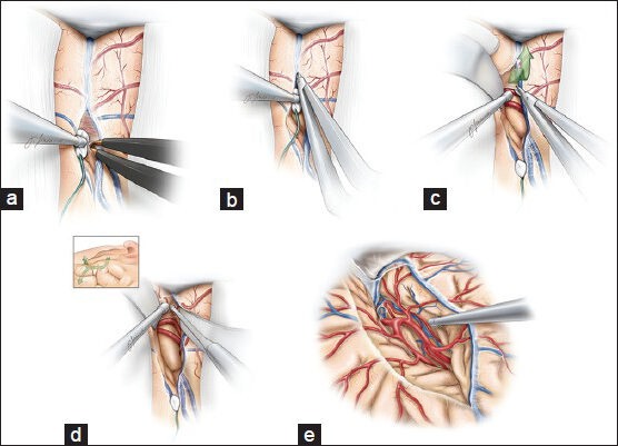
Figure 1.

The dissection of the Sylvian fissure is carried out in a deep to superficial fashion (a-d). A wide opening of the fissure is necessary to obviate the need to fixed rigid retractors. The M1 segment is identified as it curves medially (d-e). Care is taken to minimize retraction on the frontotemporal opercula and manipulation of all the vessels coursing through the fissure. This figure is from The Neurosurgical Atlas, ©Aaron A. Cohen-Gadol, MD, MSc, used with permission.
