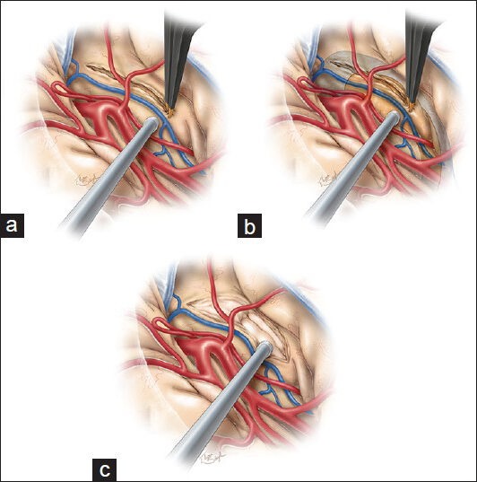
Figure 2.

Once the temporal stem has been exposed, a 10–20 mm cortical incision is made along the inferior insular sulcus. This incision begins just posterior to the temperopolar artery and lateral to the inferior parasylvian vein (a). This maneuver will allow access to the anterior temporal horn and hippocampus (b) and will create the corridor through which the amygdala and hippocampus are viewed (c). The tip of the temporal horn should be exposed to orient the surgeon regarding the adjacent anatomy. This figure is from The Neurosurgical Atlas, ©Aaron A. Cohen-Gadol, MD, MSc, used with permission.
