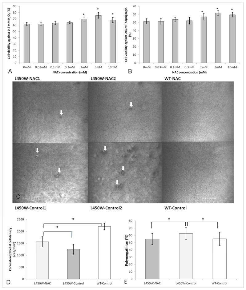
Figure 1.
Cell viability of N-acetylcysteine (NAC) treated bovine corneal endothelial cells compared to untreated control after H2O2 (A) or thapsigargin (B) conditioning (*p<0.05). Confocal microscopy of corneal endothelial cells (C) in two different mutant mice treated with NAC (L450W-NAC1/2) show fewer guttae (white arrows) and relatively normal cell size and shape compared to two different untreated mutant mice (L450W-Control1/2). Wild-type mice with (WT-NAC) and without (WT-Control) NAC treatment show normal corneal endothelium. Scale bar = 60 μm for all images (C). Mutant mice treated with NAC (L450W-NAC) show higher endothelial cell density (D) than mutant mice untreated with NAC (L450W-Control). WT mice untreated with NAC (WT-Control) are shown for comparison (D, *p<0.05). Mutant mice treated with NAC (L450W-NAC) show lower endothelial cell polymegathism (E) than mutant mice untreated with NAC (L450W-Control). WT mice untreated with NAC (WT-Control) are shown for comparison (E, *p<0.05).

