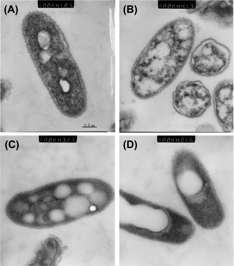
Fig. 4.
Ultrastructure of actively growing cells. (A), VBNC cells in microcosm maintained for 5 days with 200 μM copper sulfate (B), culturable cells in microcosm maintained for 5 days without copper (C), and dead cells (D) observed after ultra-thin section. Note the reduction of electron-dense granules and PHB disintegration in VBNC cells in panel B. Magnification for all 4 figures was 50,000×.

