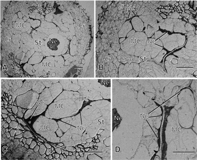
Fig. 3.
Light micrographs of root tissues of the resistant carrot line infected with the root-knot nematode at 7 weeks after inoculation, showing the formation of relatively small modified cells (MCs) around the infecting nematodes (Ne) in the stele (St) (A, C, D), and the formation of extensive necrotic layers (Nc) through the middle lamella of the modified cells and around the nematodes, demarcating the modified cells from each other and from the infecting nematodes (B, C, D). Note degenerative-looking nematodes (A, C, D). Cr: cortex. Bars = 50 μm.

