Figure 3.

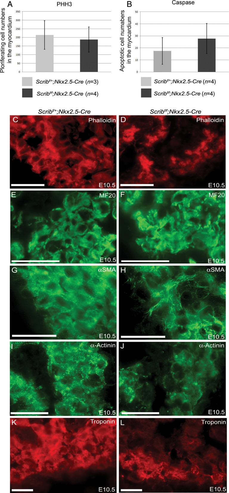
Abnormal expression of cardiomyocyte markers in Scribf/f;Nkx2.5-Cre. (A and B) Neither proliferation (pHH3) nor cell death (cleaved caspase-3) was altered in myocardium from Scribf/f;Nkx2.5-Cre ventricles compared with control littermates at E10.5. (C–L) While phalloidin and MF20 staining did not show any reproducible differences between control and mutant cardiomyocytes in the E10.5 ventricle (C–F), analysis of other markers suggested that the Scribf/f;Nkx2.5-Cre ventricles were immature in comparison with control littermates; whereas striations suggesting the presence of sarcomeres (labelled by α-SMA and α-actinin) were readily apparent in control cells, these were absent in the mutant cells (H and J). In addition, there was markedly reduced expression of cardiac troponin I (K and L) in Scribf/f;Nkx2.5-Cre trabeculae. n = 3 for all experiments. Scale bar = 20 µm.
