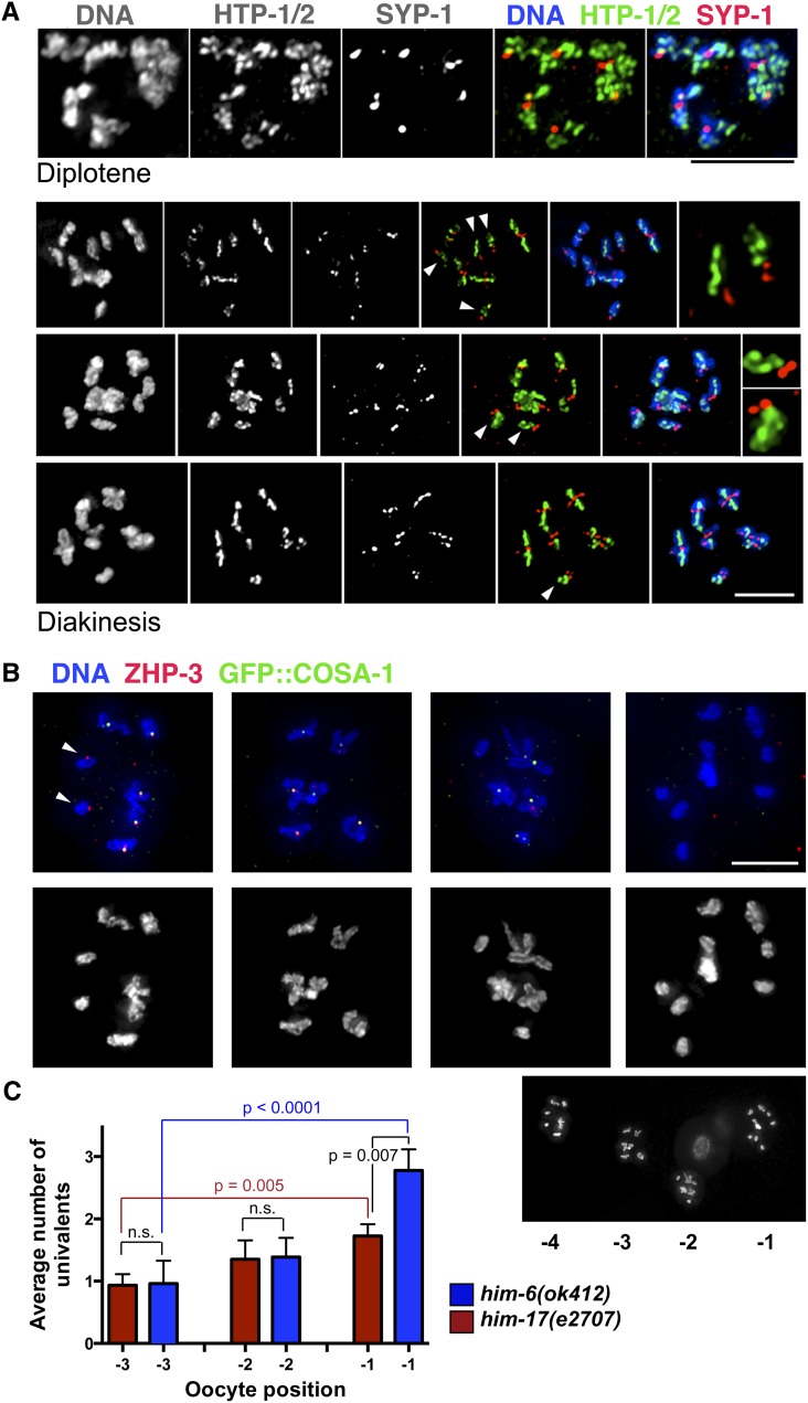
Figure 4.
Dissociation of some chromosome pairs into univalents by the end of diakinesis in him-6 mutants. (A) Diplotene and diakinesis-stage oocytes from the him-6 mutant, stained with antibodies against chromosome axis proteins HTP-1/2 and SC central region protein SYP-1. The chromosomes appear indistinguishable from wild type at the diplotene stage (Martinez-Perez et al. 2008), with the SYP-1 and HTP-1/2 proteins localizing to reciprocal domains as the chromosomes desynapse. By diakinesis, a mixture of bivalents and univalents (arrowheads) are detected; moreover, the univalents exhibit a reciprocal localization pattern for HTP-1/2 and SYP-1 that is normally associated with a crossover/chiasma. Right: For the top two diakinesis nuclei, the insets show selected univalents. (B) Full chromosome complements of individual him-6 diakinesis oocytes (from a single germ line, shown below). Oocyte nuclei from left to right were in the −4, −3, −2, and −1 positions relative to the spermatheca, with the −1 oocyte being the most mature. Arrows indicate univalents that have ZHP-3 foci, which normally mark crossover sites. Scale bars, 5μm. (C) Graph showing that the incidence of univalents in the him-6 mutant increases as oocytes progress through the diakinesis stage. The him-17(e2707) mutant, in which a decrease in DSB formation is responsible for the reduction in crossovers/chiasmata, is used as a control; in the him-17(e2707) control, there is a modest increase in the number of univalents scored during progression from the −3 to −1 position, reflecting improved ability to detect univalents as chromosome compaction increases during oocyte maturation. In contrast, there is a larger and more significant increase in univalents observed between the −3 and −1 oocytes in the him-6 mutant, suggesting the presence of more persistent connections that eventually dissociate (only a subset of P-values is depicted; see text). Error bars indicate SEM. Numbers of nuclei scored: him-17, n = 95; him-6, n = 83.

