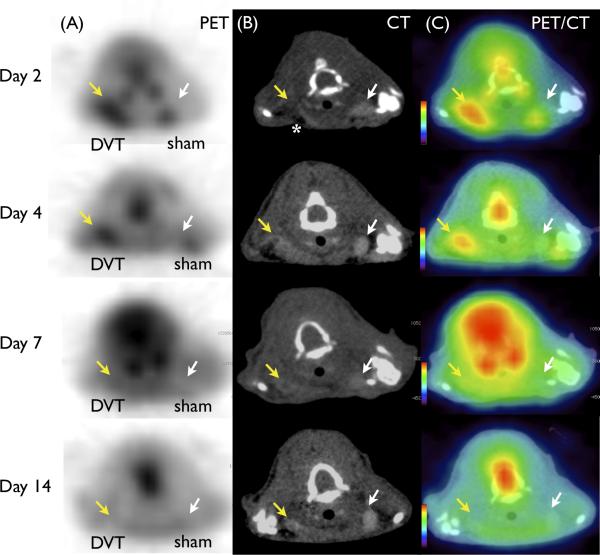
Figure 2.
FDG-PET enhances acute DVT in mice. Representative images of (A) FDG-PET (B) contrast-enhanced CT venography, and (C) fused PET/CT at various timepoints. DVT (yellow arrow) induced filling defects in contrast CT venography not seen in the contralateral sham-operated vein (white arrow). Surgery-induced air artifact (dark signal) is observed anterior to each vein on CT (white asterisk). FDG-PET DVT signal was elevated in acute DVT, and diminished over time. Mild surgery-induced inflammation at wound healing region was observed in the sham surgery left jugular vein region at day 2.

